
Imaging Services

Our imaging services encompass a wide range of diagnostic and therapeutic procedures that utilize various imaging modalities to visualize internal body structures, organs, and tissues. These services play a crucial role in the diagnosis, treatment planning, and monitoring of various medical conditions.
Here's an overview of pathology services offered in a cancer hospital:
- X-ray (Radiography):
- General X-rays: Used to diagnose conditions affecting the chest, abdomen, and extremities.
- Fluoroscopy: Real-time X-ray imaging used for procedures such as barium studies, joint injections, and gastrointestinal exams.
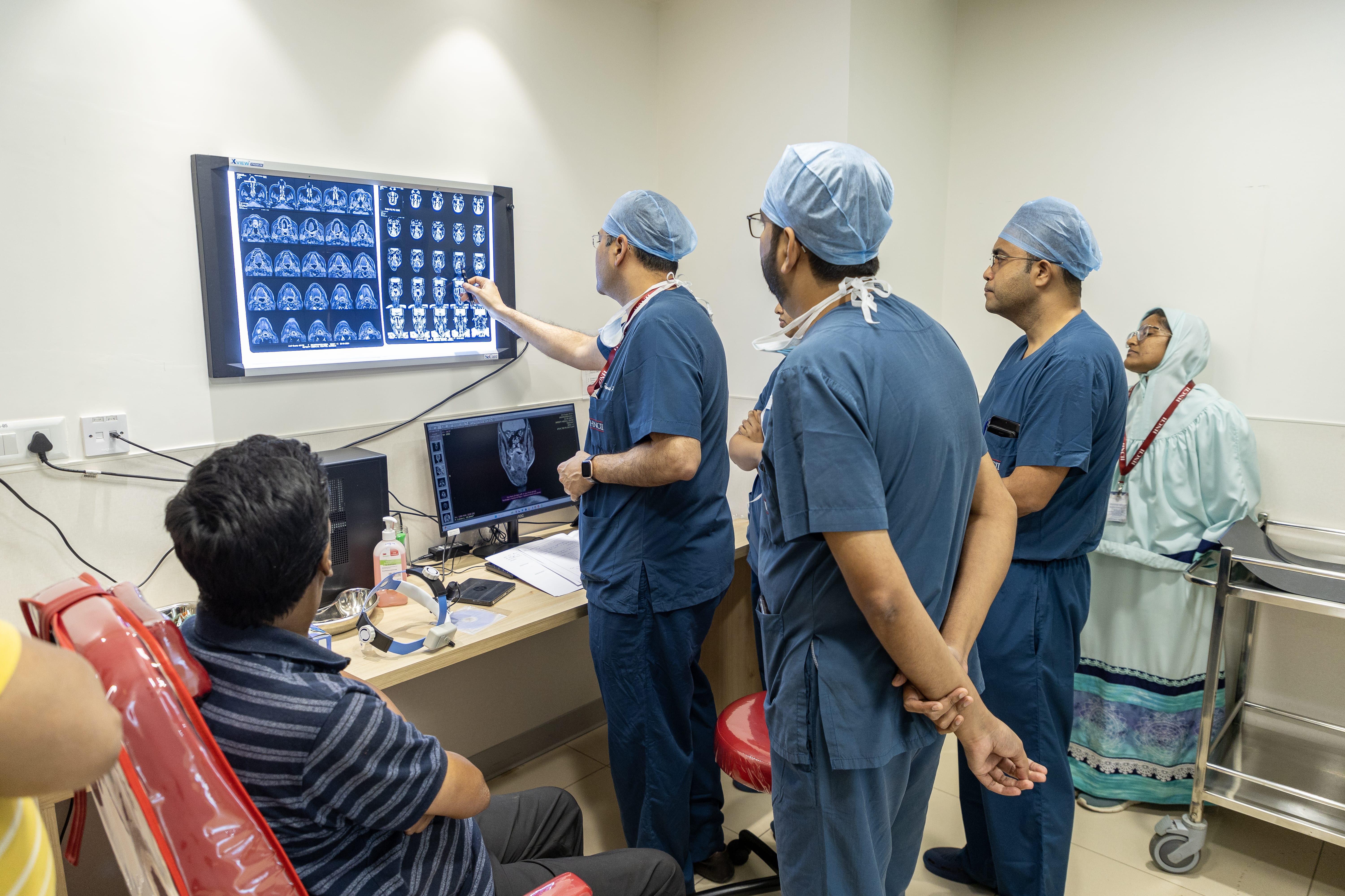
- Computed Tomography (CT):
- CT Scans: Provide detailed cross-sectional images of the body, useful for diagnosing cancers.
- Ultrasound Imaging:
- Diagnostic Ultrasound: Non-invasive imaging technique used to visualize organs, blood vessels, and to guide biopsies or needle aspirations (FNAC).
- Doppler Ultrasound: Measures blood flow and detects abnormalities in blood vessels, aiding in the diagnosis of conditions such as deep vein thrombosis (DVT) or carotid artery stenosis.
- Nuclear Medicine:
- Positron Emission Tomography (PET): Imaging technique used to detect metabolic activity in tissues, helping diagnose cancer.
- Single Photon Emission Computed Tomography (SPECT): Similar to PET but uses different tracers to visualize organ function and blood flow.
- Interventional Radiology (IR):
- Minimally Invasive Procedures: IR procedures involve using imaging guidance (such as fluoroscopy, CT, or ultrasound) to perform interventions such as tumor ablation, embolization, and biopsy.
- Mammography:
- Breast Imaging: Mammography is a specialized X-ray technique used for breast cancer screening and diagnosis. Digital mammography and tomosynthesis (3D mammography) are advanced techniques that offer improved detection and diagnostic accuracy.
- 2D Echo:
- Echocardiography, a non-invasive imaging technique used to visualize the structure and function of the heart in real-time using ultrasound technology.
- 2D echo services play a crucial role in the diagnosis, evaluation, and management of cardiovascular diseases, providing clinicians with detailed information on cardiac structure and function in a safe and non-invasive manner.
.jpg)